
3D fluid-dynamic ovarian cancer model resembling systemic drug administration for efficacy assay
Article Sidebar
Main Article Content
Abstract
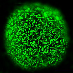
Recently, 3D in vitro cancer models have become important alternatives to animal tests for establishing the efficacy of anticancer treatments. In this work, 3D SKOV-3 cell-laden alginate hydrogels were established as ovarian tumor models and cultured within a fluid-dynamic bioreactor (MIVO®) device able to mimic the capillary flow dynamics feeding the tumor. Cisplatin efficacy tests were performed within the device over time and compared with (i) the in vitro culture under static conditions and (ii) a xenograft mouse model with SKOV-3 cells, by monitoring and measuring cell proliferation or tumor regression, respectively, over time. After one week of treatment with 10 μM cisplatin, viability of cells within the 3D hydrogels cultured under static conditions remained above 80%. In contrast, the viability of cells within the 3D hydrogels cultured within dynamic MIVO® decreased by up to 50%, and very few proliferating Ki67-positive cells were observed through immunostaining. Analysis of drug diffusion, confirmed by computational analysis, explained that these results are due to different cisplatin diffusion mechanisms in the two culture conditions. Interestingly, the outcome of the drug efficacy test in the xenograft model was about 44% of tumor regression after 5 weeks, as predicted in a shorter time in the fluid-dynamic in vitro tests carried out in the MIVO® device. These results indicate that the in vivo-like dynamic environment provided by the MIVO® device allows to better model the 3D tumor environment and predict in vivo drug efficacy than a static in vitro model.
Article Details

This work is licensed under a Creative Commons Attribution 4.0 International License.
Articles are distributed under the terms of the Creative Commons Attribution 4.0 International license (http://creativecommons.org/licenses/by/4.0/), which permits unrestricted use, distribution and reproduction in any medium, provided the original work is appropriately cited (CC-BY). Copyright on any article in ALTEX is retained by the author(s).
Abbott, A. (2003). Biology’s new dimension. Nature 424, 870-872. doi:10.1038/424870a
Amsden, B. (1998). Solute diffusion within hydrogels. Mechanisms and models. Macromolecules 31, 8382-8395. doi:10.1021/ma980765f
Aston, W. J., Hope, D. E., Nowak, A. K. et al. (2017). A systematic investigation of the maximum tolerated dose of cytotoxic chemotherapy with and without supportive care in mice. BMC Cancer 17, 684. doi:10.1186/s12885-017-3677-7
Brancato, V., Oliveira, J. M., Correlo, V. M. et al. (2020). Could 3D models of cancer enhance drug screening? Biomaterials 232, 119744. doi:10.1016/j.biomaterials.2019.119744
Burleson, K. M., Casey, R. C., Skubitz, K. M. et al. (2004). Ovarian carcinoma ascites spheroids adhere to extracellular matrix components and mesothelial cell monolayers. Gynecol Oncol 93, 170-181. doi:10.1016/j.ygyno.2003.12.034
Cavo, M., Caria, M., Pulsoni, I. et al. (2018). A new cell-laden 3D Alginate-Matrigel hydrogel resembles human breast cancer cell malignant morphology, spread and invasion capability observed “in vivo.” Sci Rep 8, 5333. doi:10.1038/s41598-018-23250-4
Chitcholtan, K., Asselin, E., Parent, S. et al. (2013). Differences in growth properties of endometrial cancer in three dimensional (3D) culture and 2D cell monolayer. Exp Cell Res 319, 75-87. doi:10.1016/j.yexcr.2012.09.012
Clevers, H. (2016). Modeling development and disease with organoids. Cell 165, 1586-1597. doi:10.1016/j.cell.2016.05.082
Curtin, C., Nolan, J. C., Conlon, R. et al. (2018). A physiologically relevant 3D collagen-based scaffold-neuroblastoma cell system exhibits chemosensitivity similar to orthotopic xenograft models. Acta Biomater 70, 84-97. doi:10.1016/j.actbio.2018.02.004
Daniel, A. B., Strickland, J., Allen, D. et al. (2018). International regulatory requirements for skin sensitization testing. Regul Toxicol Pharmacol 95, 52-65. doi:10.1016/j.yrtph.2018.03.003
Das, V., Bruzzese, F., Konečný, P. et al. (2015). Pathophysiologically relevant in vitro tumor models for drug screening. Drug Discov Today 20, 848-855. doi:10.1016/j.drudis.2015.04.004
Dutta, R. C. and Dutta, A. K. (2009). Cell-interactive 3D-scaffold; advances and applications. Biotechnol Adv 27, 334-339. doi:10.1016/j.biotechadv.2009.02.002
Fang, Y. and Eglen, R. M. (2017). Three-dimensional cell cultures in drug discovery and development. SLAS Discov 22, 456-472. doi:10.1177/1087057117696795
Fatehullah, A., Tan, S. H. and Barker, N. (2016). Organoids as an in vitro model of human development and disease. Nat Cell Biol 18, 246-254. doi:10.1038/ncb3312
Faul, F., Erdfelder, E., Lang, A.-G. et al. (2007). G*Power 3: A flexible statistical power analysis program for the social, behavioral, and biomedical sciences. Behav Res Methods 39, 175-191. doi:10.3758/bf03193146
Gao, Y., Shan, N., Zhao, C. et al. (2015). LY2109761 enhances cisplatin antitumor activity in ovarian cancer cells. Int J Clin Exp Pathol 8, 4923-4932.
Herter, S., Morra, L., Schlenker, R. et al. (2017). A novel three-dimensional heterotypic spheroid model for the assessment of the activity of cancer immunotherapy agents. Cancer Immunol Immunother 66, 129-140. doi:10.1007/s00262-016-1927-1
Hoarau-Véchot, J., Rafii, A., Touboul, C. et al. (2018). Halfway between 2D and animal models: Are 3D cultures the ideal tool to study cancer-microenvironment interactions? Int J Mol Sci 19, 181. doi:10.3390/ijms19010181
Huber, J. M., Amann, A., Koeck, S. et al. (2016). Evaluation of assays for drug efficacy in a three-dimensional model of the lung. J Cancer Res Clin Oncol 142, 1955-1966. doi:10.1007/s00432-016-2198-0
Hutmacher, D. W., Loessner, D., Rizzi, S. et al. (2010). Can tissue engineering concepts advance tumor biology research? Trends Biotechnol 28, 125-133. doi:10.1016/j.tibtech.2009.12.001
Ip, C. K. M., Li, S. S., Tang, M. Y. H. et al. (2016). Stemness and chemoresistance in epithelial ovarian carcinoma cells under shear stress. Sci Rep 6, 26788. doi:10.1038/srep26788
Jiguet Jiglaire, C., Baeza-Kallee, N., Denicolaï, E. et al. (2014). Ex vivo cultures of glioblastoma in three-dimensional hydrogel maintain the original tumor growth behavior and are suitable for preclinical drug and radiation sensitivity screening. Exp Cell Res 321, 99-108. doi:10.1016/j.yexcr.2013.12.010
Kaushik, K. H., Sripuram, V. K., Bedada, S. et al. (2010). A simple and sensitive validated HPLC method for quantitative determination of cisplatin in human plasma. Clin Res Regul Aff 27, 1-6. doi:10.3109/10601330903490462
Khurana, A. and Godugu, C. (2018). Alginate-based three-dimensional in vitro tumor models: A better alternative to current two-dimensional cell culture models. In B. Rehm and M. Moradali (eds.), Alginates and Their Biomedical Applications (157-183). Springer Series in Biomaterials Science and Engineering. Volume 11. Singapore: Springer. doi:10.1007/978-981-10-6910-9_6
Kretzschmar, K. and Clevers, H. (2016). Organoids: Modeling development and the stem cell niche in a dish. Dev Cell 38, 590-600. doi:10.1016/j.devcel.2016.08.014
LaBarbera, D. V., Reid, B. G. and Yoo, B. H. (2012). The multicellular tumor spheroid model for high-throughput cancer drug discovery. Expert Opin Drug Discov 7, 819-830. doi:10.1517/17460441.2012.708334
Lancaster, M. A. and Knoblich, J. A. (2014). Generation of cerebral organoids from human pluripotent stem cells. Nat Protoc 9, 2329-2340. doi:10.1038/nprot.2014.158
Lhuissier, E., Bazille, C., Aury-Landas, J. et al. (2017). Identification of an easy to use 3D culture model to investigate invasion and anticancer drug response in chondrosarcomas. BMC Cancer 17, 490. doi:10.1186/s12885-017-3478-z
Liu, X., Weaver, E. M. and Hummon, A. B. (2013). Evaluation of therapeutics in three-dimensional cell culture systems by MALDI imaging mass spectrometry. Anal Chem 85, 6295-6302. doi:10.1021/ac400519c
Loessner, D., Stok, K. S., Lutolf, M. P. et al. (2010). Bioengineered 3D platform to explore cell-ECM interactions and drug resistance of epithelial ovarian cancer cells. Biomaterials 31, 8494-8506. doi:10.1016/j.biomaterials.2010.07.064
Longati, P., Jia, X., Eimer, J. et al. (2013). 3D pancreatic carcinoma spheroids induce a matrix-rich, chemoresistant phenotype offering a better model for drug testing. BMC Cancer 13, 95. doi:10.1186/1471-2407-13-95
Lowe, K. A., Chia, V. M., Taylor, A. et al. (2013). An international assessment of ovarian cancer incidence and mortality. Gynecol Oncol 130, 107-114. doi:10.1016/j.ygyno.2013.03.026
Markovitz-Bishitz, Y., Tauber, Y., Afrimzon, E. et al. (2010). A polymer microstructure array for the formation, culturing, and high throughput drug screening of breast cancer spheroids. Biomaterials 31, 8436-8444. doi:10.1016/j.biomaterials.2010.07.050
Marrella, A., Lagazzo, A., Barberis, F. et al. (2017). Enhanced mechanical performances and bioactivity of cell laden-graphene oxide/alginate hydrogels open new scenario for articular tissue engineering applications. Carbon N Y 115, 608-616. doi:10.1016/j.carbon.2017.01.037
Marrella, A., Giannoni, P., Pulsoni, I. et al. (2018). Topographical features of graphene-oxide-functionalized substrates modulate cancer and healthy cell adhesion based on the cell tissue of origin. ACS Appl Mater Interfaces 10, 41978-41985. doi:10.1021/acsami.8b15036
Marrella, A., Dondero, A., Aiello, M. et al. (2019). Cell-laden hydrogel as a clinical-relevant 3D model for analyzing neuroblastoma growth, immunophenotype, and susceptibility to therapies. Front Immunol 10, 1876. doi:10.3389/fimmu.2019.01876
Marrella, A., Buratti, P., Markus, J. et al. (2020). In vitro demonstration of intestinal absorption mechanisms of different sugars using 3D organotypic tissues in a fluidic device. ALTEX 37, 255-264. doi:10.14573/altex.1908311
Modok, S., Scott, R., Alderden, R. A. et al. (2007). Transport kinetics of four- and six-coordinate platinum compounds in the multicell layer tumour model. Br J Cancer 97, 194-200. doi:10.1038/sj.bjc.6603854
Nicodemus, G. D. and Bryant, S. J. (2008). Cell encapsulation in biodegradable hydrogels for tissue engineering applications. Tissue Eng Part B Rev 14, 149-165. doi:10.1089/ten.teb.2007.0332
Nyga, A., Cheema, U. and Loizidou, M. (2011). 3D tumour models: Novel in vitro approaches to cancer studies. J Cell Commun Signal 5, 239-248. doi:10.1007/s12079-011-0132-4
Panczyk, T., Jagusiak, A., Pastorin, G. et al. (2013). Molecular dynamics study of cisplatin release from carbon nanotubes capped by magnetic nanoparticles. J Phys Chem C 117, 17327-17336. doi:10.1021/jp405593u
Raghavan, S., Ward, M. R., Rowley, K. R. et al. (2015). Formation of stable small cell number three-dimensional ovarian cancer spheroids using hanging drop arrays for preclinical drug sensitivity assays. Gynecol Oncol 138, 181-189. doi:10.1016/j.ygyno.2015.04.014
Sankaranarayanan, R. and Ferlay, J. (2006). Worldwide burden of gynaecological cancer: The size of the problem. Best Pract Res Clin Obstet Gynaecol 20, 207-225. doi:10.1016/j.bpobgyn.2005.10.007
Shamir, E. R. and Ewald, A. J. (2014). Three-dimensional organotypic culture: Experimental models of mammalian biology and disease. Nat Rev Mol Cell Biol 15, 647-664. doi:10.1038/nrm3873
Shin, Y., Han, S., Jeon, J. S. et al. (2012). Microfluidic assay for simultaneous culture of multiple cell types on surfaces or within hydrogels. Nat Protoc 7, 1247-1259. doi:10.1038/nprot.2012.051
Siegel, R., Naishadham, D. and Jemal, A. (2012). Cancer statistics for Hispanics/Latinos, 2012. CA Cancer J Clin 62, 283-298. doi:10.3322/caac.21153
Simian, M. and Bissell, M. J. (2017). Organoids: A historical perspective of thinking in three dimensions. J Cell Biol 216, 31-40. doi:10.1083/jcb.201610056
Stock, K., Estrada, M. F., Vidic, S. et al. (2016). Capturing tumor complexity in vitro: Comparative analysis of 2D and 3D tumor models for drug discovery. Sci Rep 6, 28951. doi:10.1038/srep28951
Suggitt, M. and Bibby, M. C. (2005). 50 Years of preclinical anticancer drug screening: Empirical to target-driven approaches. Clin Cancer Res 11, 971-981.
Talukdar, S. and Kundu, S. C. (2012). A non-mulberry silk fibroin protein based 3D in vitro tumor model for evaluation of anticancer drug activity. Adv Funct Mater 22, 4778-4788. doi:10.1002/adfm.201200375
Tang, Y. J., Sun, Z. L., Wu, W. G. et al. (2015). Inhibitor of signal transducer and activator of transcription 3 (STAT3) suppresses ovarian cancer growth, migration and invasion and enhances the effect of cisplatin in vitro. Genet Mol Res 14, 2450-2460. doi:10.4238/2015.March.30.3
Tezcan, S., Özdemir, F., Turhal, S. et al. (2013). High performance liquid chromatographic determination of free cisplatin in different cancer types. Der Pharma Chem 5, 169-174.
Thoma, C. R., Zimmermann, M., Agarkova, I. et al. (2014). 3D cell culture systems modeling tumor growth determinants in cancer target discovery. Adv Drug Deliv Rev 69-70, 29-41. doi:10.1016/j.addr.2014.03.001
Tiwari, A., Krishna, N. S., Nanda, K. et al. (2005). Benign prostatic hyperplasia: An insight into current investigational medical therapies. Expert Opin Investig Drugs 14, 1359-1372. doi:10.1517/13543784.14.11.1359
Trujillo-de Santiago, G., Flores-Garza, B. G., Tavares-Negrete, J. A. et al. (2019). The tumor-on-chip: Recent advances in the development of microfluidic systems to recapitulate the physiology of solid tumors. Materials (Basel) 12, 2945. doi:10.3390/ma12182945
Weaver, V. M. and Roskelley, C. D. (1997). Extracellular matrix: The central regulator of cell and tissue homeostasis. Trends Cell Biol 7, 40-42. doi:10.1016/S0962-8924(97)30078-6
Weeber, F., Ooft, S. N., Dijkstra, K. K. et al. (2017). Tumor organoids as a pre-clinical cancer model for drug discovery. Cell Chem Biol 24, 1092-1100. doi:10.1016/j.chembiol.2017.06.012
Xu, X., Farach-Carson, M. C. and Jia, X. (2014). Three-dimensional in vitro tumor models for cancer research and drug evaluation. Biotechnol Adv 32, 1256-1268. doi:10.1016/j.biotechadv.2014.07.009